Comprehensive Eye Exams
Out of the five human senses, vision has often been regarded as the most vital. Without vision, perceiving the physical world around us becomes an impairment. This can have profound implications on quality of life, safety, health, morbidity, and even mortality. In addition, the eyes are often described as windows to our health. In fact, the eye is the only part of the human body where blood vessels and nerves can be directly visualized. This allows evaluation for and even diagnosis of a host of other diseases.
The eye is evaluated by performing a comprehensive eye examination. The American Academy of Ophthalmology (AAO) recommends that all healthy adults should have a complete eye exam preferably once in their 20s and twice in their 30s. After the age of 40yrs, many adults require exams every 1-2yrs as their reading vision becomes weaker. Adults who are 60yrs and older should have their eyes examined every year.
Regardless of age, one needs to see an ophthalmologist in the following scenarios;
- Infection, injury, allergies, reduced or changed vision, pain or discomfort, flashes or floaters in the vision, or any new eye symptoms
- Having an eye disease that needs regular follow up, like glaucoma, dry eye disease, blepharitis, strabismus, macular degeneration, epiretinal membranes, vitreous floaters etc.
- Medical conditions like diabetes, uncontrolled hypertension, thyroid disease, multiple sclerosis, neurological diseases and many autoimmune diseases can affect the eyes and screening exams should be done at least once a year.
- Family history of certain diseases, like glaucoma, melanoma or macular degeneration, may require annual screenings
- If you take medications that can affect vision (eg hydroxychloroquine, amiodarone or prednisone), talk to your ophthalmologist about how often the eyes should be screened.
- If you wear glasses or contact lenses, it is recommended that the health of the eyes be checked annually. These vision exams are normally performed by our optometrists.
The eyes should be screened for age-related diseases such as;
- Cataracts
- Glaucoma
- Age-related macular degeneration
- Retinal tears and detachments
- Dry eye disease
What should I expect during a comprehensive eye exam?
The eye exam is comfortable and spans between 45 and 90minutes. A technician will likely perform some of the more routine aspects of the exam, but the critical portions of viewing and assessing the eye are performed by your eye doctor. The exam includes the following;
History
A basic history and review of your current and past medical conditions, medications, allergies and surgeries, if any. Many systemic diseases and medications have potential side-effects or complications in the eye. It is important to bring a complete list of your current medication and details of prior and current medical conditions to your appointment for our doctors to review.
Vision and Refraction
The visual acuity will be checked with and/or without glasses/contact lenses.
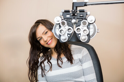
A refraction will be done to determine the correct glasses prescription for you. A refraction also helps the doctor determine what your best visual acuity is which reflects
the health of the eye. This is done primarily by having you look through a machine
called a phoropter, which will allow different lenses to be presented to determine which
combination gives you the best vision. Sometimes, the doctor will perform this manually using hand-held lenses. All our offices are equipped with a dedicated optical department and experienced opticians that specialize in the highest quality lenses and frames for your perusal.
Contact lens fittings
If you wear contact lenses, it is recommended to get your eyes checked once a year. Our optometrists offer new contact lens fittings and rechecks for all ages, from children to adolescents to adults. They are able to fit a range of contact lens options. Soft contact lenses are available in daily, biweekly, and monthly disposables, toric contact lenses to correct astigmatism, and multifocal contact lenses to correct near and distance vision. Our optometrists also have the expertise to fit Rigid gas-permeable (RGP) lenses or scleral lenses for regular use and therapeutically to treat disorders like keratoconus or irregular astigmatism. MiSight soft lenses are available for treatment of rapidly progressive myopia in children.
Pupils, Peripheral vision and Ocular movements
A light is shone into the pupils check how they respond to the light. You may be asked to identify a target in various positions in your peripheral visual field to ensure they are intact. The alignment and movement of the eyes is assessed by having you fixate on a point light source. If there is double vision, it can be measured using prisms.
Eye pressure check
This is a screening test for glaucoma and is painless. Anesthetic eyedrops are instilled in the eyes before an instrument is used to check the eye pressure.
Dilation

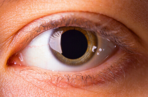
Eye drops are instilled to allow the pupils to dilate fully so that the internal structures of the eye can be visualized. The dilation can make the reading, and sometimes distance, vision blurry for 4-12h.
While many people are able to drive with their eyes dilated, it is recommended that you have someone who can drive you home if you are not comfortable doing so.
Slit-lamp examination
The slit lamp is a machine used by your eye doctor to evaluate the eye using enhanced lighting and magnification. Through the slit lamp, fine structures in the eye like the cornea, iris, lens, vitreous humor, optic nerve, blood vessels and retina can be visualized in great detail. Your doctor will have you place your chin on a chin rest while using a narrow beam of light to look into your eye. This exam is easy and comfortable.
Indirect Ophthalmoscopy
In order for the peripheral retina to be assessed, the eyes must be first dilated and then visualized using indirect ophthalmoscopy. Your doctor will have you sit back in your exam chair and look into the eyes using a headset with a light and a magnifying lens. This is the only way the peripheral retina can be be seen, and is necessary to check for diseases like diabetes, retinal tears or detachments, and problems with blood circulation among others.
Additional testing
Depending on the findings during your eye exam, your doctor may perform additional testing. A full range of ancillary testing is offered in our offices. These include;
- Tear osmolarity and Inflammadry
These involve sampling the tear film to evaluate dry eye disease - Lipiview
Imaging the meibomian glands and tear film for ocular surface disease - Corneal topography
Mapping the surface of the cornea - Ocular Coherence Tomography (OCT)
Scanning the retina and/or nerve tissue with ultrasound to evaluate diseases like glaucoma and retinal disease - Visual field testing
A 15min test in which the peripheral visual field is mapped by having you press a button every time you see a light stimulus at various points in your visual field. This is used to evaluate glaucoma and other neurological diseases. - Fundus photography
Pictures of the entire retina to document disease - Gonioscopy
The doctor looks into the drainage angles within the eye with a special lens to evaluate for types of glaucoma and diabetes - Pachymetry
Measurement of corneal thickness using a hand held instrument that gently taps on the eye - Color vision and Stereo-acuity testing
Simple interactive tests to determine color vision and binocular functioning - A scan and B-scan ultrasonography
Utilizing ultrasound to measure and visualize the inside of the eyeball


